| |
Linearity of Submicro Labeling
|
| |
| In order to assess the linearity of the 3DNA labeling method on arrays, parallel RNA titration experiments were performed on spotted oligonucleotide and cDNA arrays. The oligo arrays consisted of approximately 70mer oligonucleotides spotted on poly-L-lysine (The Center for Applied Genomics, NJ). These oligonucleotides were derived from mouse low, medium, and high level expressed genes. The cDNA arrays used for these studies were 13K Mouse Chips from Agilent. For both experiments the same mouse total RNA samples were reverse transcribed in both channels following the Genisphere Submicro EX, Appendix A protocol. A 2-step hybridization procedure was used for both array types |
| |
| Mouse Oligo Array Hybridization: |
| |
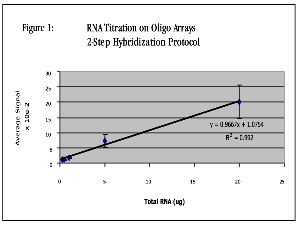
| The total RNA was titrated from 0.2µg to 20µg in both channels. The cDNA was hybridized overnight (16 hours) in Genisphere Vial 6 hybridization buffer at 55°C. On the following morning the arrays were washed for 15 minutes with 2X SSC/0.2% SDS at 42°C, 10 minutes in 2X SSC at room temperature, and 10 minutes in 0.2X SSC at room temperature as described in the Submicro protocol. The arrays were dried and incubated in a 65°C hybridization oven for 20 minutes to prewarm the array. 2.5 microliters each of the Cy3 and Cy5 Capture Reagents were combined in Genisphere Vial 6 hybridization buffer. This mixture was scaled up so that there would be enough for all of the arrays in the experiment. The 3DNA hybridization mixture was added to each array and the arrays were hybridized for 2.5 hours at 65°C. The arrays were washed as described above except that the first wash temperature was 65°C instead of 42°C. The arrays were scanned on an Axon Genepix 4000B scanner at PMT settings that yielded a balanced signal with no saturated features on the 20µg total RNA input arrays. These settings were left constant for all subsequent scans. The data was extracted using Genepix 3.0 software. The average Cy3 and Cy5 signals were calculated for each RNA input data set. The Cy3 and Cy5 data were then averaged and plotted against the input RNA (Figure 1). |
| |
 |
| |
| The average signal was transformed for plotting purposes by dividing each intensity by 100. Since 20µg of total RNA resulted in an average signal of 2000, a slope of 1.000 on the plot of intensity (x100) versus input RNA would indicate a perfect dose response (i.e. 2-fold increase in RNA results in a 2-fold increase in average signal intensity). The slope of the titration line was calculated to be 0.9667 and had a correlation of 0.992. |
| |
| |
| Mouse 13K Agilent Array Hybridization: |
| |
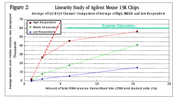
| The total RNA was titrated from 1.0µg to 16.3µg in both channels. The cDNA was hybridized overnight (16 hours) in Agilent Deposition Hybridization Buffer at 65°C. On the following morning the arrays were washed for 5 minutes with 0.5X SSC, 0.01% SDS (Agilent Wash Solution 1) and then for 5 minutes in 0.06X SSC at room temperature. The arrays were dried and incubated in a 55°C hybridization oven for 20 minutes to prewarm the array. 2.5 microliters each of the Cy3 and Cy5 capture reagents were combined in Genisphere 2X Vial 7 hybridization buffer. This mixture was scaled up so that there would be enough for all of the arrays in the experiment. The 3DNA hybridization mixture was added to each array and the arrays were hybridized for 3 hours at 53°C. The arrays were washed for 15 minutes with 2X SSC/0.2%SDS at 42°C, 10 minutes in 2X SSC at room temperature, and 10 minutes in 0.2X SSC at room temperature as described in the Submicro protocol. The arrays were scanned on an Axon Genepix 4000B scanner at PMT settings that yielded a balanced signal between channels and some saturated features on the 16.3µg input RNA arrays. These settings were left constant for all subsequent scans. The data was extracted using Genepix 3.0 software. The data were subdivided between low, medium and high level expressers and the average of 10 genes from each category was calculated for each channel. The Cy3 and Cy5 data for each category were then averaged and plotted against the input RNA (Figure 2). As indicated by the resulting plots of the low and medium level expressers, the response is linear over the range of the RNA titrated. Also a 2.5 fold increase in RNA input results in nearly a 2.5 fold increase in signal. When analyzing the results of the high level expressers the signal intensities reached a maximum at about 5-7.5µg of input total RNA. The intensity at this point was defined by the maximum measurable intensity of the scanner, indicated as "Scanner Saturation" on the plot. If the scanner's dynamic range had been greater, then the plot for the high level expressers would have extended linearly, as is indicated by the red dashed line in Figure 2. |
| |
 |
| |
| |
 |
