Appendix B. cDNA Gels
|
| Required Materials |
 |
cDNA sample (approximately 1ug RNA equivalent) |
|
Vertical Electrophoresis System (Novex model no. E19001-XCELL II MiniCell) |
|
10% TBE-Urea Gel (Novex Cat. No. EC6875BOX) |
|
1X TBE running buffer |
|
4x denaturing load dye (50% formamide, 50mM Tris, 1 mM EDTA, 0.01% Bromophenol Blue/Xylene Cyanol) |
|
SybrGold Nucleic Acid Gel Stain (Molecular Probes Cat. No. S-11494) |
|
Transilluminator (VWR Scientific model no. VWR LM20E) |
|
Power Supply |
|
|
 |
|
| Step 1: Preparation of Gel |
|
|
| 1. |
Remove wrapping and comb from a 1mm Novex 10% acrylamide/TBE urea gel and rinse wells with RGDD water. Remove lower strip. |
| 2. |
Assemble Novex vertical gel apparatus and insert gel with large plate facing out. Insert balance if only running one gel and insert wedge. Tighten. |
| 3. |
Heat 1 L TBE to approximately 65oC. |
| 4. |
Fill inside and outside section of gel box to top with warmed 1X TBE. Make sure there are no bubbles under gel or in wells. |
| 5. |
Wash wells several times with running buffer (using syringe). |
| |
|
|
| Step 2: Preparation of Samples |
|
|
| 1. |
Combine cDNA (1 ug RNA equivalent) from reverse transcription reaction (after neutralization) with 3 ul 4X denaturing load dye. Example: take 1/5th of a 5 ug total RNA reaction. Bring volume to 12 ul with water. |
| 2. |
Mix and briefly microfuge samples. Heat to 70oC 3 minutes. |
| |
|
|
| Step 3: Running Gel |
|
|
| 1. |
Rinse wells with running buffer just prior to loading samples. |
| 2. |
Load entire sample prepared above. |
| 3. |
Place cover on top of gel, attach electrodes to Power Supply and turn on power. |
| 4. |
Run at 150V for 45 min. |
| |
|
|
| Step 4: Gel Staining, Imaging |
|
|
| 1. |
Make up a solution of SybrGold Gel Stain (1:10,000 SybrGold in running buffer). |
| 2. |
Disassemble gel unit. Transfer gel to SybrGold Gel Stain solution. Incubate 40 minutes (in dark, light stirring). |
| 3. |
Illuminate gel on transilluminator (300nm excitation). |
| 4. |
Capture image (digital or film). |
| |
|
|
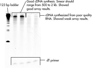
| Interpreting Gel Results: |
|
|
| |
High-quality cDNA will appear as a smear between roughly 500 and 2kb. |
|
| |
| a. |
OD 260/280 ratio will be between 1.9 and 2.1. |
| b. |
On an agarose gel, total plant and mammalian RNA will be represented as two sharp, bright bands. For mammalian RNA, the bands will be at ~ 4.5 kb and ~ 1.9 kb, representing the 28S and 18S ribosomal sub-units, respectively. Please refer to the image below. |
|
| |
 |
| |
 |
|
|
