Appendix A. RNA Gels
|
| Required Materials |
 |
Total RNA sample (approximately 1 ug) |
|
Agarose- LE (Ambion Cat. No. 9040) |
|
NorthernMax Formaldehyde Load Dye (Ambion Cat. No. 8550G) |
|
NorthernMax 10X Denaturing Gel Buffer (Ambion Cat. No. 8676) |
|
NorthernMax 10X MOPS Gel Running Buffer (Ambion Cat. No. 8671) |
|
Horizontal Gel Electrophoresis System (Life Technologies Cat. No. 11068-012) |
|
Power Supply |
|
SybrGold Nucleic Acid Gel Stain (Molecular Probes Cat. No. S-11494) |
|
Transilluminator (VWR Scientific model no. VWR LM20E) |
|
RNaseZAP (Ambion Cat. No. 9780) |
|
|
 |
|
| Step 1: Preparation of Gel |
|
|
| 1. |
Clean all equipment (pipettors, glassware, gel unit etc.) with RNaseZAP to ensure an RNase-free environment. |
| 2. |
Assemble Horizontal Gel Electrophoresis System. |
| 3. |
Dissolve 1 g agarose in 90ml RNase-free water. Allow to cool to 50-60°C. |
| 4. |
Add 10 ml 10X Denaturing Gel Buffer (do this in a ventilating hood), mix thorougly. |
| 5. |
Pour the gel to between 0.6cm and 1cm in thickness. Allow to solidify at room temperature or 4°C. |
| 6. |
Dilute 10X MOPS Gel Running Buffer to 1X with nuclease-free water. Cover gel in running buffer. |
| |
|
|
| Step 2: Preparation of Sample RNA |
|
|
| 1. |
In a 1.5 ml microfuge tube, combine RNA (approx. 1ug) with 3ul NorthernMax Formaldehyde Load Dye. Bring volume to 10ul with nuclease-free water. |
| 2. |
Heat sample to 65oC 15 minutes. |
| |
|
|
| Step 3: Running Gel |
|
|
| 1. |
Load the RNA sample into wells. |
| 2. |
Attach electrode wires from gel unit to power supply. |
| 3. |
Run the gel at approximately 5V/cm for 60-90 minutes. |
| |
|
|
| Step 4: Gel Staining, Imaging |
|
|
| 1. |
Make up a solution of SybrGold Gel Stain (1:10,000 SybrGold in running buffer). |
| 2. |
Disassemble gel unit. Transfer gel to SybrGold Gel Stain solution. Incubate 40 minutes (in dark, light stirring). |
| 3. |
Illuminate gel on transilluminator (300nm excitation). |
| 4. |
Capture image (digital or film). |
| |
|
|
| Interpreting Gel Results: |
|
|
| |
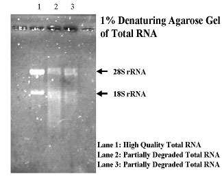
High-quality RNA will have the following characteristics: |
|
| |
| a. |
OD 260/280 ratio will be between 1.9 and 2.1. |
| b. |
On an agarose gel, total plant and mammalian RNA will be represented as two sharp, bright bands. For mammalian RNA, the bands will be at ~ 4.5 kb and ~ 1.9 kb, representing the 28S and 18S ribosomal sub-units, respectively. Please refer to the image below. |
|
| |
 |
| |
|
|
|